Peptide molecular weight can be worked out from the sequence alone: add the mass of each amino-acid residue, then add one water. This guide shows the arithmetic for a short public sequence, explains how the isoelectric point (pI) is estimated, and lists why the weight on a certificate of analysis (COA) can differ from a calculator.
Quick answer: How do you calculate a peptide’s molecular weight? Add the residue mass of every amino acid in the sequence, then add the mass of one water molecule. GHK works out to 340.186 (monoisotopic) and 340.38 (average), in daltons or g/mol.
What a residue mass is
IUPAC’s amino-acid nomenclature rules explain the idea. When two or more amino acids combine to form a peptide, the elements of water are removed. What remains of each amino acid is called a residue. Our guide to peptide bond formation covers that reaction.
So a residue mass is the mass of an amino acid minus one water. A chain of residues still has a free amino end and a free carboxyl end, and those ends add back one water in total. The Expasy Compute pI/Mw tool states the rule in its documentation: molecular weight is the sum of the average masses of the amino acids in the protein plus the average mass of one water molecule.
Average vs monoisotopic mass
Most elements occur as more than one isotope. NIST’s isotope tables show the split. Carbon is 98.93% carbon-12 and 1.07% carbon-13. Hydrogen, nitrogen and oxygen also carry small amounts of heavier isotopes.
- Average mass uses standard atomic weights, which average over that natural mix. The CIAAW abridged table gives hydrogen 1.0080, carbon 12.011, nitrogen 14.007 and oxygen 15.999.
- Monoisotopic mass uses the exact mass of the most abundant isotope of each element. From NIST: hydrogen-1 is 1.00782503223, carbon-12 is exactly 12, nitrogen-14 is 14.00307400443 and oxygen-16 is 15.99491461957.
Water shows the difference. Two hydrogen-1 atoms plus one oxygen-16 give 18.01056. Two average hydrogens plus one average oxygen give about 18.015.
Size matters too. By our arithmetic from the NIST abundances, about 83% of GHK molecules contain only the lightest isotopes. Senko and colleagues noted that in large biomolecules the many atoms make the all-monoisotopic species unlikely.
Worked example: GHK
GHK is the tripeptide glycyl-histidyl-lysine. PubChem lists it as CID 73587, formula C14H24N6O4. The residue masses below are from Expasy’s table of mass values.
Molecular weight of GHK, step by step
| Component | Monoisotopic | Average |
|---|---|---|
| Glycine residue (G) | 57.02146 | 57.0519 |
| Histidine residue (H) | 137.05891 | 137.1411 |
| Lysine residue (K) | 128.09496 | 128.1741 |
| Sum of residues | 322.17533 | 322.3671 |
| Plus one water (H2O) | 18.01056 | 18.01524 |
| GHK, total | 340.18589 | 340.3823 |
| PubChem, for comparison | 340.18590327 | 340.38 |
The sums match PubChem’s computed values to four decimal places (monoisotopic) and two (average); the last-digit gap is rounding. A free peptide calculator that runs this arithmetic for any sequence is coming to the site.
Modifications that change the mass
The rule above assumes a linear chain with free ends. Common changes shift the total by known amounts. The values below are monoisotopic, from the Unimod database:
- Disulfide bond. Each cysteine in a disulfide bridge loses one hydrogen (−1.007825), so one bridge lowers the mass by about 2.016.
- C-terminal amide. Replacing the terminal acid with an amide changes the mass by −0.984016.
- N-terminal acetyl. Adding an acetyl group adds 42.010565.
- Metal complexes. PubChem’s record for the copper complex of GHK, prezatide copper, has formula C14H23CuN6O4+ and a weight of 402.92. That is GHK plus one copper (63.546) minus one hydrogen. A separate record, prezatide copper acetate (C32H54CuN12O12), lists 862.4. By atom count, that formula is two GHK units, one copper and two acetates. Our GHK-Cu guide covers the complex itself.
Isoelectric point and net charge
Kozlowski’s 2016 paper on the Isoelectric Point Calculator defines the isoelectric point as the pH at which a protein’s net charge is zero, and notes that the Henderson-Hasselbalch equation can be used to calculate charge at a given pH.
Each ionizable group has a pKa. For a basic group (the N-terminal amine, Lys, Arg, His), the positive charge at a given pH is 1 / (1 + 10(pH − pKa)). For an acidic group (the C-terminal carboxyl, Asp, Glu, Cys, Tyr), the negative charge is 1 / (1 + 10(pKa − pH)). Sum the groups for net charge. The pI is the pH where that sum reaches zero.
GHK has four ionizable groups: the N-terminal amine, the His side chain, the Lys side chain and the C-terminal carboxyl. We ran the arithmetic with three pKa sets tabulated by Kozlowski.
GHK net charge at pH 7 and estimated pI, by pKa set
| pKa set | N-term / His / Lys / C-term pKa | Net charge at pH 7 | Estimated pI |
|---|---|---|---|
| EMBOSS | 8.6 / 6.5 / 10.8 / 3.6 | +1.22 | 9.70 |
| Lehninger | 9.69 / 6.0 / 10.5 / 2.34 | +1.09 | 10.10 |
| IPC_peptide | 9.564 / 6.018 / 10.517 / 2.383 | +1.09 | 10.04 |
All three sets put GHK at about +1 at pH 7: the N-terminal amine and Lys are mostly protonated, His only partly, and the carboxyl end is negative. The estimated pI moves by 0.4 units between sets.
Why tools give different pI values
Kozlowski writes that pKa estimates differ with the experimental setup, and that more than 600 different pKa values have been reported. Expasy uses values from Bjellqvist and colleagues, measured in immobilized pH gradient gels with 9.2 M and 9.8 M urea, and its tool does not account for post-translational modifications. Kozlowski adds that pI is easier to predict for short peptides than for proteins.
Why the molecular weight on a COA can differ
A certificate can print a number that does not match a calculator, with nothing wrong with the sample:
- Mass type. Monoisotopic and average figures differ, by about 0.2 for GHK and more for longer chains.
- Charged ions. Fenn and colleagues described electrospray spectra as runs of peaks whose ions differ by one charge from their neighbors. A printed MS value can belong to a charged ion, not the neutral molecule.
- Salt form. A 2020 review by Sikora and colleagues notes that peptides are cleaved and purified with trifluoroacetic acid (TFA), so cationic peptides come out as TFA salts, and that most peptide drugs are supplied as acetate salts. Bachem’s quality-control guide says the acid is bound to the free N-terminus and to basic side chains such as Arg, Lys and His.
- Metal or modification. A copper complex or a terminal cap changes the formula, as shown above.
As arithmetic only, GHK with two acetic acid counter-ions (60.052 each) weighs 460.49, of which the peptide is 73.9%. With two TFA counter-ions (114.022 each) it weighs 568.43, and the peptide is 59.9%. Bachem adds that peptides rich in basic amino acids should be expected to have a low net peptide content, even when very pure, because of salt formation.
So purity and peptide content are separate numbers. Bachem defines net peptide content as the percentage of peptide relative to non-peptide material, mostly counter-ions and moisture, and states that it is not the same as purity. Purity is typically read from HPLC with UV detection at 210–220 nm, as the main peak’s share of the total peak area. Our guides to HPLC purity and reading a COA show where each figure sits on a certificate.
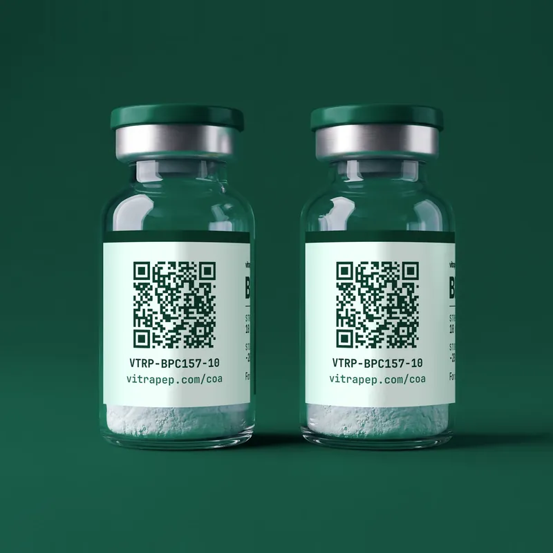
Vitrapep Research Catalog
Research peptides supplied as laboratory reference compounds, each lot with a third-party certificate of analysis. For laboratory research use only, not for human or veterinary use.
Frequently asked questions
How do you calculate the molecular weight of a peptide?
Add the residue mass of each amino acid in the sequence, then add one water: 18.01056 for monoisotopic mass or about 18.015 for average mass. Adjust for disulfides, terminal caps, metals or salts.
What is the isoelectric point of a peptide?
The pH at which its net charge is zero. It is estimated from pKa values for the termini and the charged side chains, and the estimate depends on which pKa set is used.
Why doesn’t the weight on a COA match a calculator?
The certificate may use the other mass type, report a charged ion, or describe a salt or metal complex. Counter-ions such as acetate or TFA add weight that is not peptide.
What to know now
- The rule: molecular weight is the sum of residue masses plus one water.
- Two mass types: monoisotopic uses the most abundant isotopes; average uses standard atomic weights.
- GHK: 340.186 monoisotopic, 340.38 average, formula C14H24N6O4.
- Modifications: a disulfide bridge removes about 2.016; amidation and acetylation have fixed shifts.
- pI: the pH of zero net charge, estimated from pKa values that vary by source.
- COA figures: salts, metals and moisture explain most gaps; net peptide content is not purity.